Mass Spectrometry Analysis for Peptides: A How-To Guide
Table of Contents
- How Peptide Mass Spectrometry Analysis Works
- Peptide Sample Preparation for Mass Spectrometry
- Peptide Mapping Mass Spectrometry for Protein Characterization
- Peptide Identification by Mass Spectrometry: From Spectra to Sequence
- Peptide Mass Spectrum Interpretation: Reading m/z and Fragment Ions
- Troubleshooting Common Peptide Mass Spectrometry Problems
- Frequently Asked Questions
Last Updated: October 14, 2026
How Peptide Mass Spectrometry Analysis Works
Mass spectrometry analysis for peptides is the process of ionizing peptide molecules, separating them by mass-to-charge ratio, and reading the resulting spectrum to determine what a sample contains. At FROPeptides, we rely on this technique to verify every batch we ship, and it is the same method research labs use to characterize proteins, confirm sequences, and catch impurities.
The core idea is simple. A peptide is weighed, broken apart in a controlled way, and the fragments are counted. The pattern of fragments tells you the sequence.
From Sample to Spectrum: The Core Workflow
Every peptide mass spectrometry run follows the same path:
- Prepare the sample and digest proteins into peptides
- Separate peptides by liquid chromatography
- Ionize the peptides so they carry a charge
- Measure the mass-to-charge ratio (m/z) of each ion
- Fragment selected ions and measure the pieces
- Match the data against a sequence database
Each step feeds the next. A weak step one ruins everything downstream.
The most common cause of a failed run is not the instrument. It is a dirty sample. Salt, detergent, and polymer residue suppress ionization and flatten your signal before the mass analyzer ever sees it.
Peptide Sample Preparation for Mass Spectrometry
Peptide sample preparation for mass spectrometry decides the quality of everything that follows. Poor prep produces noisy spectra, low sequence coverage, and false negatives.
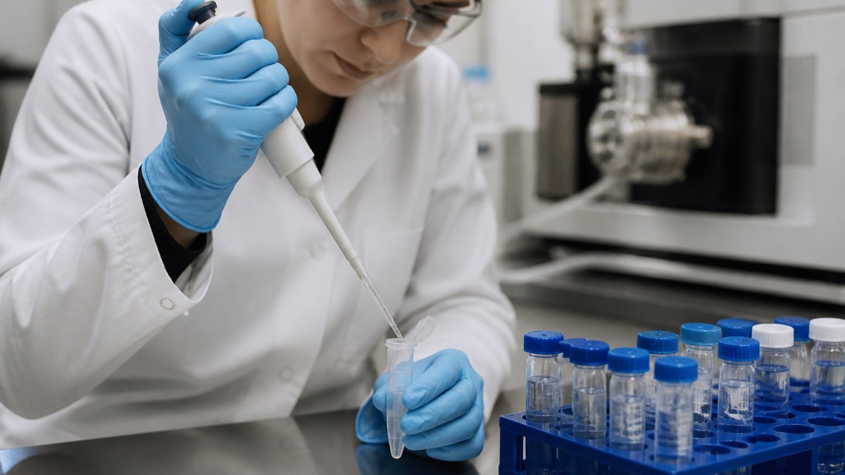
Start with clean handling. Keep tubes capped, work on ice where the protocol calls for it, and use fresh buffers.
Reduction, Alkylation, and Enzymatic Digestion
For protein samples, the standard path is:
- Reduction breaks disulfide bonds with a reagent such as dithiothreitol
- Alkylation caps the freed thiols with iodoacetamide to stop them re-forming
- Enzymatic digestion cuts the protein into peptides, usually with trypsin
- Cleanup removes salts and reagents before the instrument
Trypsin is the workhorse because it cuts after lysine and arginine, giving peptides in a mass range that instruments handle well. Let it digest overnight at a controlled temperature for the most complete cut.
Skipping the alkylation step leaves disulfide bonds free to re-form. The result is missing peptides, gaps in sequence coverage, and a digest that looks incomplete for no obvious reason.
Peptide Mapping Mass Spectrometry for Protein Characterization
Peptide mapping mass spectrometry confirms that a protein matches its expected sequence. You digest the protein, run the peptides, and compare the observed masses against the masses predicted from the known sequence.
This is the standard method for protein characterization in biologics work. It catches sequence variants, unexpected modifications, and truncations that a simple mass check would miss.
Two numbers matter most:
| Metric | What It Tells You | Why It Matters |
|---|---|---|
| Sequence coverage | Percent of the protein you actually observed | Low coverage means blind spots |
| Mass accuracy | How close measured mass is to expected | Tight accuracy confirms identity |
High sequence coverage plus tight mass accuracy gives you confidence the protein is what you think it is.
Peptide Identification by Mass Spectrometry: From Spectra to Sequence
Peptide identification by mass spectrometry works by matching experimental fragment patterns to theoretical ones. The instrument breaks each peptide in predictable places, and the resulting fragment ions form a fingerprint unique to that sequence.
Database Search Versus De Novo Sequencing
Two approaches dominate:
- Database search compares your spectra to a library of predicted sequences. It is faster, more sensitive, and the default for most proteomics work.
- De novo sequencing reads the sequence directly from the fragment ladder. It is slower but handles novel peptides, unusual organisms, and sequences absent from any database.
Software such as Mascot, SEQUEST, and open-source tools like MS-GF+ handle the matching. Each search engine has strengths, Mascot and SEQUEST are long-established in core facilities, while MS-GF+ and similar open-source tools are favored for reproducibility and cost. Many labs run two engines and compare results.
Setting Confidence Thresholds
A confident peptide identification needs more than one matching fragment ion. Single-fragment matches are noise until proven otherwise. The practical controls are:
- False discovery rate (FDR): the expected proportion of incorrect identifications among accepted results. A 1% FDR is the common target for publication-grade work; 5% is sometimes used for exploratory runs.
- Decoy database: a reversed or shuffled version of the target database used to estimate the FDR empirically. If your search does not use a decoy, your FDR number is a guess.
- Score threshold: the minimum match score a peptide must reach. Set it based on the decoy distribution, not on a round number.
- Peptide-spectrum match (PSM) count: the number of spectra supporting a given peptide. More supporting spectra mean more confidence.
A loose threshold inflates your hit count with junk. A tight threshold throws away real peptides. The right setting depends on whether you are discovering or confirming.
From Identification to Quantification
Identification answers “what is in the sample.” Quantification answers “how much.” These are different questions and require different methods.
Label-free quantification compares signal intensity or spectral counts across runs.
Labeled quantification uses stable-isotope tags or metabolic labels to compare samples within a single run. It is more precise but more expensive and requires additional sample handling.
Targeted quantification uses selected reaction monitoring or parallel reaction monitoring to measure specific peptides with high sensitivity and a wide dynamic range.
Calibration, Accuracy, and Limits
Every quantitative method needs calibration. The core performance metrics are:
- Accuracy: how close the measured value is to the true value. Assessed with certified reference standards or spiked-in heavy peptides.
- Precision: how close repeated measurements are to each other. Reported as a coefficient of variation across replicates.
- Limit of detection (LOD): the lowest amount that can be reliably distinguished from background.
- Limit of quantification (LOQ): the lowest amount that can be measured with acceptable accuracy and precision.
- Linearity: the range over which signal response is proportional to amount.
A method that has not been characterized for these metrics is a method you cannot defend in a report. For regulated work, validation follows established guidance from the U.S. Food and Drug Administration on bioanalytical method validation, which sets expectations for accuracy, precision, selectivity, and stability.
Reporting Results
A defensible report states the search engine and version, the database and its version, the FDR threshold, the number of decoy hits, the sequence coverage, and the mass accuracy. Without these, a reader cannot judge whether the identification is real. Include the raw spectra for any peptide that carries a conclusion.
A confident peptide identification needs more than one matching fragment ion, and a confident quantitative result needs calibration, precision data, and a stated limit of quantification. Identification and quantification are separate claims that require separate evidence.
Peptide Mass Spectrum Interpretation: Reading m/z and Fragment Ions
Peptide mass spectrum interpretation starts with the precursor ion. That is the intact peptide’s mass-to-charge ratio, recorded before fragmentation. It tells you the peptide’s overall mass.
Then the instrument fragments the precursor and records the product ions. In collision-induced dissociation, the peptide breaks along the backbone, producing a ladder of b and y ions.
A few practical rules:
- The mass difference between adjacent y ions reveals one amino acid
- The most intense peaks are not always the most informative
- Charge state changes the m/z, so always check the charge before calculating mass
Interpretation is a skill built by repetition. Run known standards first so you learn what a clean spectrum looks like.
Troubleshooting Common Peptide Mass Spectrometry Problems
Most failures trace back to a short list of causes. Work through them in order before blaming the instrument. The decision tree below is organized by the symptom you actually see on screen, because that is how problems present in practice.
Symptom-First Decision Tree
No signal at all. Check the spray first, a stable electrospray plume should be visible at the emitter tip.
Weak signal with high background. This is almost always a sample cleanliness problem, not an instrument problem. Salt, detergent, and polymer residue suppress ionization.
Missing peptides or gaps in sequence coverage. Incomplete digestion is the usual cause.
Poor fragmentation with few product ions. Collision energy is the first lever.
| Problem | Likely Cause | Fix |
|---|---|---|
| No signal at all | Ionization failure or clogged column | Check spray, replace column |
| Weak signal | Sample too dilute or salty | Concentrate and clean up sample |
| Missing peptides | Incomplete digestion | Extend digest time, add enzyme |
| High background | Contamination | Use fresh solvents, clean glassware |
| Poor fragmentation | Wrong collision energy | Tune energy for peptide size |
| Unexpected mass | Wrong charge state or missed modification | Verify charge, check for oxidation |
Contamination Control
Keratins from skin, dust, and clothing are the most common contaminants in a peptide lab. Wear gloves, keep tubes closed, and clean the source regularly. Polymer residue from plasticware and detergent residue from glassware are the next most common offenders. A periodic blank run is the cheapest diagnostic you can build into your workflow.
Matching the Method to the Sample and the Goal
Troubleshooting is easier when you start with the right method. Match the tool to the sample type and the question you are asking.
| Sample Type | Goal | Recommended Approach |
|---|---|---|
| Intact protein | Confirm molecular weight, detect truncations | Intact-mass analysis, high-resolution analyzer |
| Digested protein | Confirm sequence, find variants | Peptide mapping with LC-MS/MS |
| Complex mixture | Deep sequence coverage, discovery | Bottom-up LC-MS/MS with data-dependent acquisition |
| Synthetic peptide, crude | Confirm identity and purity | MALDI or LC-MS, fast check |
| Synthetic peptide, purified | Verify sequence and modifications | LC-MS/MS with targeted fragmentation |
| Unknown or novel peptide | Determine sequence without a database | De novo sequencing from fragment ladder |
Use LC-MS/MS for complex mixtures and deep sequence coverage. Use MALDI for fast, simple checks on clean samples. Use an Orbitrap analyzer when you need high resolution and accurate mass. For targeted work where you already know what you are looking for, a triple quadrupole in selected reaction monitoring mode gives the best sensitivity and dynamic range.
Before you troubleshoot the instrument, run a known standard. If the standard looks clean, the problem is in your sample. If the standard also looks bad, the problem is in the system. This one step saves hours of guessing.
Sourcing clean starting material removes a whole class of problems before they start. FROPeptides tests every batch with multi-stage HPLC and mass spectrometry, and ships with a Certificate of Analysis so you can trace purity before you run a single sample.
Frequently Asked Questions
How is mass spectrometry used for peptide identification?
Peptide mass spectrometry measures the mass-to-charge ratio (m/z) of intact peptides and their fragment ions. In bottom-up proteomics, proteins are digested into peptides, separated by liquid chromatography, ionized by electrospray ionization (ESI), and analyzed in a mass analyzer such as an Orbitrap. Tandem mass spectrometry (MS/MS) fragments the precursor ion, and the resulting product ion spectrum is matched to a sequence database to identify the peptide and its parent protein.
How do you prepare a peptide sample for mass spectrometry?
Start by desalting the sample, since salts and detergents suppress ionization. For protein analysis, reduce disulfide bonds with dithiothreitol, alkylate cysteines with iodoacetamide, then digest with trypsin at 37°C overnight. For synthetic peptides, dissolve the lyophilized powder in a compatible solvent such as 0.1% formic acid in water or acetonitrile. Keep samples cold, avoid repeated freeze-thaw cycles, and use low-binding tubes to prevent peptide loss before LC-MS/MS injection.
How do you interpret a peptide mass spectrum?
Begin with the precursor ion’s m/z and charge state to calculate the peptide’s molecular mass. In an MS/MS spectrum, look for fragment ions (b-ions and y-ions) that differ by the mass of individual amino acids. A series of y-ions or b-ions spaced by residue masses allows you to read the sequence from one end. Software such as Mascot, SEQUEST, or MaxQuant automates this by comparing observed fragments against theoretical spectra from a protein database.
What is the difference between peptide mapping and tandem mass spectrometry?
Peptide mapping uses enzymatic digestion followed by LC-MS to confirm a protein’s identity and detect modifications by comparing the observed peptide masses to the expected masses for that sequence. Tandem mass spectrometry (MS/MS) goes further by fragmenting each peptide and reading its sequence directly. Peptide mapping is common for batch-to-batch consistency testing of biologics, while MS/MS is used when you need sequence-level confirmation or to locate a specific post-translational modification.
Peptide analysis only works as well as the material you put into it. If your starting peptides carry hidden impurities, no amount of instrument tuning will rescue the data. FROPeptides supplies high-purity, lab-verified research peptides and lyophilized coenzymes, each backed by multi-stage HPLC testing, mass spectrometry verification, and batch-level Certificate of Analysis documentation. Get started with FROPeptides and run every protocol on material you can trace from vial to result.