Signs of Chemical Degradation in Vials: A Lab Guide
Table of Contents
- Introduction to Chemical Degradation in Vials
- Visual Signs of Chemical Degradation in Vials
- What Peptide Vial Crystals Actually Mean
- Reconstituted Peptide Stability: What Changes After Mixing
- Peptide Degradation Analysis Mass Spectrometry and Other Methods
- Peptide Storage Best Practices to Prevent Degradation
- Interpreting Results: When to Use or Discard a Vial
- Conclusion: Protecting Your Research Through Early Detection
- Frequently Asked Questions
Last Updated: October 7, 2026
Introduction to Chemical Degradation in Vials
Chemical degradation is the breakdown of a compound’s molecular structure over time, driven by heat, light, oxygen, or moisture. For anyone handling research peptides and lyophilized coenzymes, spotting the early signs of chemical degradation in vials is the difference between usable data and a wasted experiment. At FROPeptides, we verify every batch through multi-stage HPLC testing and mass spectrometry.
The signs fall into two groups: what you can see, and what you need an instrument to confirm. Most researchers catch the visible ones and miss the rest.
Below, we break down each warning sign, explain what it means, and show you how to decide whether a vial is still good.
Visual Signs of Chemical Degradation in Vials
Visual inspection is the fastest first check, and it costs nothing. Hold the vial against a white background under bright, diffused light. Then compare it to a fresh reference vial from the same batch if you have one.
Color Changes and Discoloration
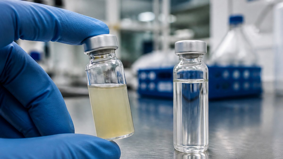
A shift in color is often the first visible sign. A peptide that should be white or off-white turning yellow, brown, or amber points to oxidation or breakdown of the compound.
Watch for these changes:
- Yellowing of a previously white powder
- Darkening at the edges of the cake
- Uneven color across the vial
- A tint that deepens over weeks in storage
Some color shift is normal for certain compounds. The problem is a change from the baseline you recorded when the vial arrived.
Cloudiness, Particles, and Precipitates
Cloudiness in a reconstituted solution almost always means trouble. A clear solution that turns hazy, or one with floating particles, has likely lost integrity.
Check for:
- Fine haze that will not clear after mixing
- Visible particles or flakes
- Sediment settled at the bottom
- A film coating the glass wall
Any of these means the compound has come out of solution. Discard it.
What Peptide Vial Crystals Actually Mean
Peptide vial crystals are solid formations that appear when a compound comes out of solution or when a lyophilized cake collapses. They are not automatically a failure, but they always deserve a closer look.
Here is the distinction that matters. A uniform, fluffy, white cake is what a good lyophilized peptide looks like. Hard crystals, glassy shards, or a melted-looking cake signal a problem with the freeze-drying process or storage conditions.
What most guides miss is that crystals can form two ways:
- During lyophilization, if the drying cycle was off
- After reconstitution, if the solution was stored cold and the peptide dropped out
The first is a manufacturing issue. The second is a handling issue. Only the second is something you can fix by warming the vial gently and re-dissolving. If the crystals will not dissolve, the peptide has likely aggregated, and aggregation usually means loss of activity.
Reconstituted Peptide Stability: What Changes After Mixing
Reconstituted peptide stability drops sharply the moment you add solvent. A lyophilized powder can hold for months or years. Once mixed, most peptides are best used within days to a few weeks, depending on the compound.
Several factors speed up the clock:
- Warm storage temperatures
- Repeated freeze-thaw cycles
- Exposure to light
- Bacterial growth in non-sterile water
- Agitation during transport
The single biggest mistake we see is researchers mixing a full vial when they only need part of it. Mix what you will use in the next week or two. Keep the rest as powder.
Never refreeze a reconstituted peptide more than once. Each freeze-thaw cycle stresses the molecule and can trigger aggregation that no amount of gentle warming will reverse.
Store reconstituted vials cold, protected from light, and use them on a fixed schedule. Label the mix date on the vial itself.
Peptide Degradation Analysis Mass Spectrometry and Other Methods
When visual checks are not enough, analytical testing gives you a definitive answer. Peptide degradation analysis mass spectrometry confirms whether the molecule is intact by measuring its exact mass. A shift in mass reveals oxidation, truncation, or other damage.
The two workhorse methods are:
| Method | What It Detects | Best For |
|---|---|---|
| HPLC | Purity, impurities, degradation peaks | Routine batch checks |
| Mass spectrometry | Exact molecular mass, structural changes | Confirmatory testing |
| Visual inspection | Color, clarity, particles | Fast first screen |
HPLC and Mass Spectrometry for Confirmatory Testing
HPLC separates the components in a sample and shows you how much of it is intact peptide versus breakdown products. A purity peak that has dropped, or new peaks that were not there before, tells you degradation has started.
Mass spectrometry then confirms the identity of the main peak. If the measured mass matches the expected mass, the molecule is intact. If it is off by a known amount, you can often tell exactly what happened, such as an added oxygen atom from oxidation.
For background on how these methods fit into pharmaceutical quality testing, the FDA guidance on analytical procedures outlines the validation standards labs are expected to meet.
Run HPLC first to screen, then mass spec to confirm. Together they give you a full picture of vial integrity.
Peptide Storage Best Practices to Prevent Degradation
Good storage is the cheapest form of quality control. Follow these peptide storage best practices and you will avoid most degradation before it starts.
- Keep lyophilized powder frozen, typically at -20°C or colder
- Protect vials from light with amber glass or foil wrap
- Keep desiccant in the storage container to limit moisture
- Let vials reach room temperature before opening, to stop condensation
- Avoid repeated temperature swings
- Log every vial’s arrival date and storage location
Temperature control matters most. A vial that warms and cools repeatedly will degrade faster than one held steady, even at a slightly warmer set point.
Photograph each new batch next to a ruler on arrival. That baseline image makes it easy to spot color or cake changes weeks later, and it takes ten seconds per vial.
For cold-chain standards used across the industry, the USP guidance on storage and distribution is a solid reference for temperature ranges.
Interpreting Results: When to Use or Discard a Vial
The decision to use or discard comes down to matching your evidence to your tolerance for risk. A vial that fails a visual check should not go into a sensitive assay.
Use this quick framework:
- Clear solution, intact cake, no color shift: use it
- Minor color shift, otherwise clean: test before use
- Cloudy, particles, or crystals that will not dissolve: discard
- Failed HPLC or mass spec: discard
For critical work, such as assays where small purity differences affect results, always confirm with analytical testing before use. For non-critical work, a clean visual check plus proper storage is often enough.
When in doubt, discard. The cost of one vial is far lower than the cost of a failed experiment or misleading data.
Conclusion: Protecting Your Research Through Early Detection
Catching the signs of chemical degradation in vials early protects both your samples and your results. Visible changes like color shift, cloudiness, and stubborn crystals are your first alert. HPLC and mass spectrometry confirm what your eyes cannot.
FROPeptides supports this work with lab-verified peptides and lyophilized coenzymes, backed by multi-stage HPLC testing, mass spectrometry on every batch, and a Certificate of Analysis you can check before you commit. Our temperature-controlled storage and batch-level traceability help ensure every vial arrives intact.
Get started with FROPeptides and give your lab research-grade materials it can rely on.
Frequently Asked Questions
What are the most common signs of chemical degradation in a peptide vial?
The most common visual signs include discoloration, cloudiness, visible particles, and the formation of crystals or precipitates. For lyophilized powders, a change from a fluffy white cake to a shrunken, sticky, or discolored mass often indicates degradation. These changes suggest that the peptide’s chemical structure may have been altered by hydrolysis, oxidation, or aggregation. Any visible change warrants further analytical testing before the vial is used in experiments.
Are crystals in a peptide vial always a sign of degradation?
Not always. Some peptides naturally form crystals during lyophilization or when stored at certain temperatures. However, crystal formation can also indicate aggregation or precipitation caused by degradation. The key is context: if crystals appear in a previously clear solution or if they are accompanied by cloudiness or color change, degradation is likely. Confirm with analytical methods like mass spectrometry or HPLC rather than relying on appearance alone.
How can I confirm whether a peptide has degraded using mass spectrometry?
Mass spectrometry identifies the molecular weight of the peptide and any impurities. A degraded sample will show additional peaks corresponding to fragments, oxidized forms, or aggregates that differ from the expected mass. When combined with HPLC, which separates components by hydrophobicity, you get a clear picture of purity and structural integrity. This approach is standard for peptide degradation analysis and provides definitive evidence of chemical changes.
What are the best storage practices to prevent peptide degradation?
Store lyophilized peptides in a freezer at -20°C or lower, desiccated and protected from light. For reconstituted peptides, always follow the supplier’s recommendations and keep detailed records of storage conditions to maintain peptide stability.